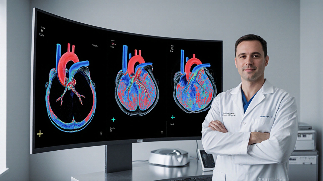
Cardiac MRI
When working with Cardiac MRI, a specialized imaging technique that creates detailed pictures of the heart’s chambers, vessels, and tissue. Also known as cardiac magnetic resonance imaging, it provides clinicians with a non‑invasive window into heart structure and function. Heart failure often reveals thickened walls or reduced pumping, while stroke risk can be assessed by spotting scar tissue or clots. Managing these conditions frequently involves cholesterol medication to keep arteries clear and beta blocker therapy to control blood pressure.
Why cardiac MRI matters for everyday care
Cardiac MRI offers unmatched detail compared to standard echo or CT scans, so doctors can spot early signs of heart failure before symptoms flare. The scan also helps identify areas at risk for stroke by highlighting scarred tissue that could trigger abnormal rhythms. When doctors prescribe cholesterol medication, they often reference MRI findings to track plaque reduction over time. Likewise, beta blockers are adjusted based on how well the heart walls move during the scan, ensuring the dose matches the patient’s actual cardiac performance.
Understanding these connections makes the image data far more than pretty pictures—it becomes a roadmap for medication choices, lifestyle tweaks, and follow‑up plans. Below you’ll find a curated set of articles that walk through buying affordable generic drugs, managing heart‑related conditions, and picking the right supplements. Use the insights from each piece to complement what a cardiac MRI can reveal about your heart’s health.
Cardiac MRI for Evaluating Hypertrophic Subaortic Stenosis
- Keith Ashcroft
- |
- |
- 13
Explore how cardiac MRI provides detailed tissue and functional insights for diagnosing and managing hypertrophic subaortic stenosis, including risk assessment, treatment planning, and future imaging advances.
View more